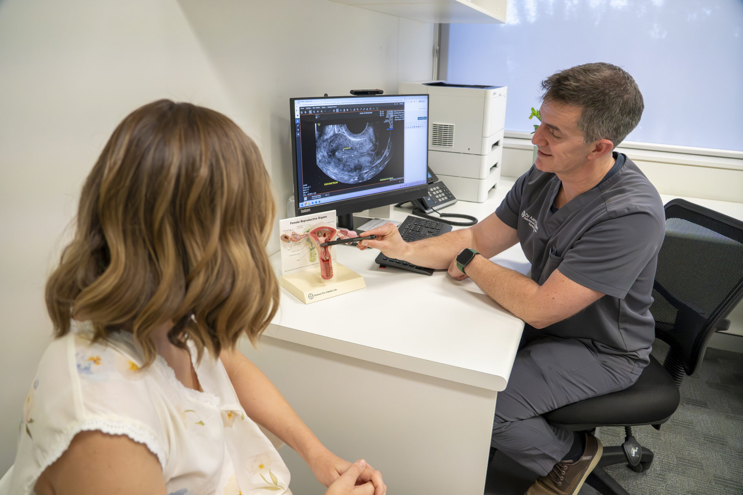
For years, the journey to an endometriosis diagnosis has been inherently frustrating, often taking a decade of navigating closed doors, dismissed pain, and vague answers. Historically, patients were told that a major, invasive surgery was the only way to truly obtain an endometriosis diagnosis.
But thankfully, medicine has evolved. Today, advanced specialised imaging can offer diagnostic clarity that matches the accuracy of surgery, completely changing how we approach, map, and treat the endometriosis symptoms that can come with the disease. If you are living with chronic pelvic pain, painful and irregular periods, or deep discomfort, you deserve a clear roadmap and clarity about what is going on. Here, we break down exactly how the modern process for endometriosis diagnosis works, and how to ensure you are getting the right tests and answers for your body.
The Key Messages You Need to Know
Before diving into the steps, there are three fundamental truths about modern endometriosis care and diagnosis that every patient and practitioner should hold onto:
- Imaging can match surgery: Specialised medical imaging, when performed by an expert operator, can have a similar sensitivity to surgery for deep and ovarian endometriosis diagnosis.
- A “normal” scan does not mean the pain isn’t real: A completely normal ultrasound or MRI cannot reliably exclude endometriosis, especially superficial peritoneal disease.
- The operator matters more than the machine: Endometriosis imaging is highly specialised and operator-dependent. Getting the right answers requires a team specifically trained in complex pelvic imaging.
The Path to Diagnosis: From First Symptoms to Clarity
The road to an accurate endometriosis diagnosis generally follows a structured, collaborative process between you, your GP, and your imaging specialists.
1. Recognising and Tracking Endometriosis Symptoms
The process begins by identifying cyclical or chronic symptoms that interfere with your quality of life. These commonly include, but are not limited to:
- Severe period pain (dysmenorrhea)
- Irregular or heavy menstrual bleeding
- Chronic pelvic pain
- Pain during or after intercourse
- Fertility challenges
- Cyclical bowel and bladder symptoms (like pain, bloating, or changes in habits during your period).
2. The GP Consultation & Detailed Referral
Your next step is a dedicated consultation with your GP. Whilst endometriosis is extremely common, your GP is best placed to ensure your symptoms are investigated appropriately and that other causes of pain are not present. They can also start discussions on treatment options for your symptoms that are right for you.
When imaging is being considered, the goal is to ensure a highly detailed referral. The more specific your referral is regarding your symptoms (e.g., noting deep pain during intercourse or severe bowel pain), the better your imaging team can provide the right diagnostic action plan to help provide clarity and a diagnosis.
3. First-Line Specialist Imaging
When endometriosis is suspected, current guidelines recommend a specialised transvaginal ultrasound as your initial diagnostic step. This ultrasound is invasive and is not possible for everyone to undertake. A transabdominal ultrasound can be undertaken but will not see subtle endometriosis, and therefore you may need to consider the following options.
The Australian Living Guideline for Endometriosis provides detailed recommendations on the steps to initiate diagnosis of endometriosis.
The 3 Diagnostic Methods: Ultrasound, MRI, Surgery
When looking for endometriosis, modern medicine utilises three primary pathways. Understanding the strengths and limitations of each helps you advocate for the right care.
1. Transvaginal Ultrasound (TVUS)
- What it is: A pelvic scan performed using a specialised probe gently placed in the vagina. It is the universal first-line test for investigating pelvic pain. Crucially, there isn’t just one type of transvaginal scan, and the exact type you receive depends entirely on your symptoms and what your GP notes on your initial referral included. If your specialist referral explicitly notes suspected endometriosis or severe chronic pain, it triggers the imaging team to perform a specialised Complex Pelvic Scan or DIE Scan instead of a basic overview.
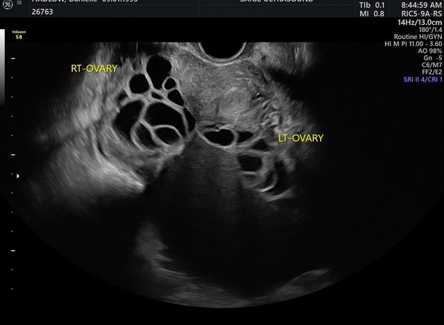
- What it finds: When your endometriosis symptoms guide us to perform a specialised protocol, it can accurately identify ovarian endometriomas (“chocolate cysts”), deep infiltrating endometriosis (DIE) nodules in the bowel or bladder, and pelvic adhesions (where organs are stuck together).
- Pros & Cons: It is highly accessible, does not expose you to radiation, and allows our team to dynamically assess organ mobility in real-time. However, because it is highly operator-dependent, it relies completely on your imaging team having advanced, dedicated training in complex gynaecological disease to accurately spot the signs.
2. Magnetic Resonance Imaging (MRI)
- What it is: An advanced, highly detailed imaging test that uses powerful magnetic fields to create structural maps of your pelvis.
- What it finds: Deep infiltrating endometriosis, complex anatomical distortion, and extra-pelvic disease (endometriosis located outside the immediate pelvic cavity).
- Pros & Cons: Excellent for pre-operative planning and viewing areas outside the reach of ultrasound. On the downside, it is more expensive, less accessible, requires a specialist referral for a Medicare (MBS) rebate, and can still miss superficial endometriosis disease. This type of diagnostic method is usually reserved for those who do not tolerate TV (trans-vaginal) ultrasound or pre-operative planning.
3. Diagnostic Laparoscopy (Surgery)
- What it is: A keyhole surgery performed under general anesthesia where a small camera is inserted through tiny abdominal incisions to look directly at the pelvic organs.
- What it finds: All forms of endometriosis, including superficial peritoneal lesions that imaging cannot see. It allows for tissue biopsies to definitively confirm the diagnosis under a microscope.
- Pros & Cons: It provides direct visualisation and allows for immediate surgical treatment/removal of the disease at the same time. However, it is an invasive surgical procedure carrying standard hospital risks, recovery time, and varies widely based on the surgeon’s specific expertise.
Note on Emerging Biomarkers
While these tests analysing blood, urine, or saliva are looking highly promising for reducing diagnostic delays in primary care, there are currently no biomarker tests with sufficient accuracy available for routine clinical use in Australia. For now, imaging and clinical evaluation remain the standard.
The 3 Types of Pelvic Ultrasound
Not all pelvic ultrasounds are created equal. When a standard imaging clinic reports a “perfectly normal scan,” it may simply mean they performed a basic check rather than a specialised endometriosis assessment.
Basic Pelvic Scan
What it involves for the patient: This scan typically begins transabdominally (on top of your tummy). You will be asked to arrive with a full bladder, which acts as a clear “window” for the ultrasound waves to see through. The sonographer will apply a warm, clear gel to your lower abdomen and move a smooth handheld wand (transducer) over the skin, applying light pressure. An internal look is often recommended to get a closer view. You will be asked to empty your bladder completely before a specialised, lubricated probe is gently inserted into the vagina. These are the base standard for assessing the pelvis and are often quick.
Best used for: Investigating heavy menstrual bleeding, checking for large uterine fibroids or massive ovarian cysts, and routine fertility or early pregnancy checks.
The core benefit: It provides an excellent, low-stress overview of general gynaecological anatomy to help rule out common, major conditions.
Complex Pelvic Scan
What it involves for the patient: Because this involves deeper, more intricate tissue issues, a transvaginal (internal) approach is required as an essential step. You will often be asked to empty your bladder completely before the scan begins, although some imaging providers will do an initial check through the abdomen first. Unlike a basic scan, this uses a specific Medicare (MBS) code that mandates a minimum scan time of 30 minutes, meaning you will be on the bed for a bit longer while the team carefully examines chronic conditions. During the scan, the specialist team will perform “dynamic mapping.” This means they will apply gentle pressure with the internal probe or their hand to your lower abdomen to see whether your organs slide freely or are stuck together. They will also check for “site-specific tenderness” to see if a particular spot corresponds to your pain.
Best used for: Investigating chronic, unexplained pelvic pain, suspected endometriosis, adenomyosis, or pelvic venous congestion.
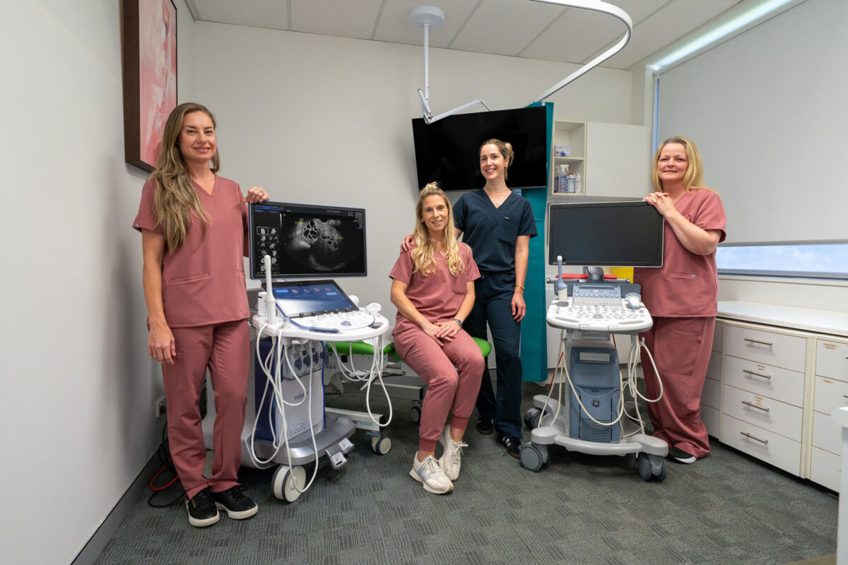
The core benefit: It utilises real-time scanning techniques (such as checking the “sliding sign”) to assess your organ mobility and determine whether your pelvic tissues are stuck together by adhesions. Conducting this type of scan requires sonographers to have undertaken specialised, advanced training, which is exactly what we provide for patients seeking an advanced ultrasound in Coffs Harbour at Saige Ultrasound.
Special Considerations: Identification of endometriosis requires specialist training and a high volume of endometriosis in the practice to ensure adequate diagnostic accuracy. The Australasian Sonographers Association (ASA) released specific Endometriosis Ultrasound Guidelinesto standardise scanning, documentation, and referral processes. The resources promote a “right scan, first time” approach, emphasising a two-tiered model to assist sonographers in both routine and advanced, endometriosis-focused pelvic ultrasound assessments.
Deep Infiltrating Endometriosis (DIE) Scan
What it involves for the patient: This is a highly comprehensive, advanced transvaginal scan that takes the most time (often between 30 and 60 minutes). To ensure the clearest possible images, you may be given specific ultrasound preparation instructions when booking, such as using a mild bowel prep or a gentle laxative the evening before to clear any gas or stool from the bowel. The scan itself is an entirely specialist-driven protocol where the internal probe is moved methodically to map out the deep spaces of the pelvis. Because the operator is systematically checking areas such as the rectum, bladder, and ligaments, you may experience localised pressure or mild discomfort as they locate and measure deep nodules. Imaging providers offering DIE scans should be following the IDEA consensus for endometriosis and report adenomyosis based on the MUSA criteria.
Best used for: Patients experiencing severe bowel or bladder symptoms tied to their cycle, advanced pre-operative surgical planning, or monitoring of known deep disease.
The core benefit: It goes far beyond standard anatomy to systematically evaluate the uterus, ovaries, Pouch of Douglas, uterosacral ligaments, bowel, rectum, bladder, ureters, and vaginal septum. It maps the exact depth, location, and surgical landscape of deep nodules that penetrate more than 5 mm beneath the tissue surface.
Note on Transvaginal Imaging:
While the thought of an internal scan can feel daunting, a transvaginal ultrasound is the definitive key to unlocking an endometriosis diagnosis; transabdominal imaging simply cannot detect deep pelvic disease. If you are navigating this journey and booking a pelvic ultrasound on the Coffs Harbour coast, we encourage you to discuss this investigation with your GP when requesting your referral so you feel fully counselled, prepared, and informed before arriving at our clinic. Your comfort is our priority; if you are unable or choose not to undergo internal imaging, our team at Saige Ultrasound will gently guide you through alternatives, such as a specialist referral for an MRI.
Whilst the upfront cost may be higher for a complex scan, the out-of-pocket costs should be approximately the same. You can view Saige Ultrasound fees for more information on out-of-pocket costs.
What If My Scan Result is “Normal”?
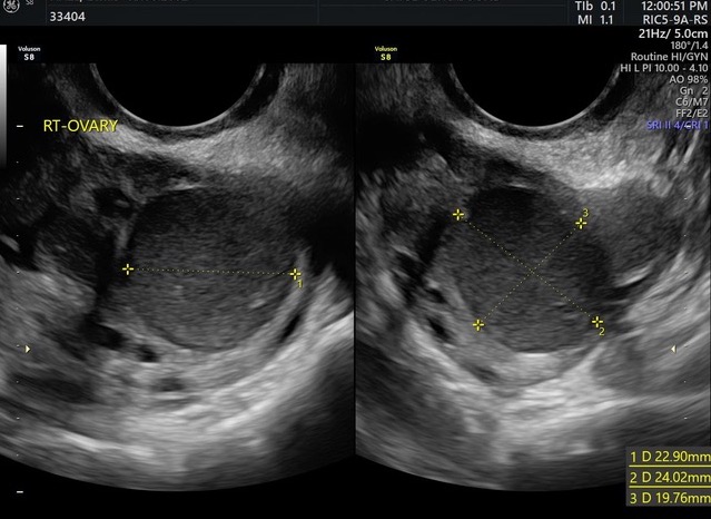
Hearing that your scan came back “normal” when you are dealing with debilitating pain can feel devastating and frustrating. But it is vital to know that a normal scan is not a blank check of health, nor does it mean the pain is in your head.
Even the most advanced ultrasounds and MRIs cannot always reliably detect superficial peritoneal endometriosis – tiny, shallow tissue spots scattered along the lining of your pelvis. If your imaging is clear but your symptoms are profoundly impacting your life, your clinical experience is what matters to us. A normal scan simply rules out deep nodules and ovarian cysts and may show “soft markers” for superficial endometriosis, allowing your medical team to confidently discuss the next steps, which may include a clinical diagnosis based on symptoms, targeted medical therapies, or a referral for a laparoscopy.
Seeking a diagnosis? How to take the first step
You do not have to live with unanswered questions or chronic, debilitating symptoms. Here is how you can proactively start your diagnostic journey with Saige Ultrasound today:
- Step 1: Track Your Symptoms. Before seeing your doctor, jot down your cycle history, noting when your pain occurs, its severity, and whether it affects your bowel, bladder, or daily activities. The more information on your symptoms that you can provide, the better. Some tools designed to help with this include QENDO app, Charli Health app and the Jean Hailes Endometriosis symptom checklist.
- Step 2: Schedule a Dedicated GP Appointment. Book a longer appointment with your doctor to discuss your symptoms more extensively. Request a detailed referral, specifically asking for a Complex Pelvic Ultrasound, and ensure they note your specific endometriosis symptoms on the request form so our team can best tailor your scan. Note: If your GP fills out your referral on a different clinic’s form, you can still bring it directly to us – read more about your referral choice options.
- Step 3: Secure Your Appointment at Saige. Contact our welcoming team at our Coffs Harbour ultrasound clinic to book your specialised assessment. Because our service is entirely specialist-led by practising Obstetricians and Gynaecologists, you can trust your care to a team with the highest level of clinical training and sensitivity.
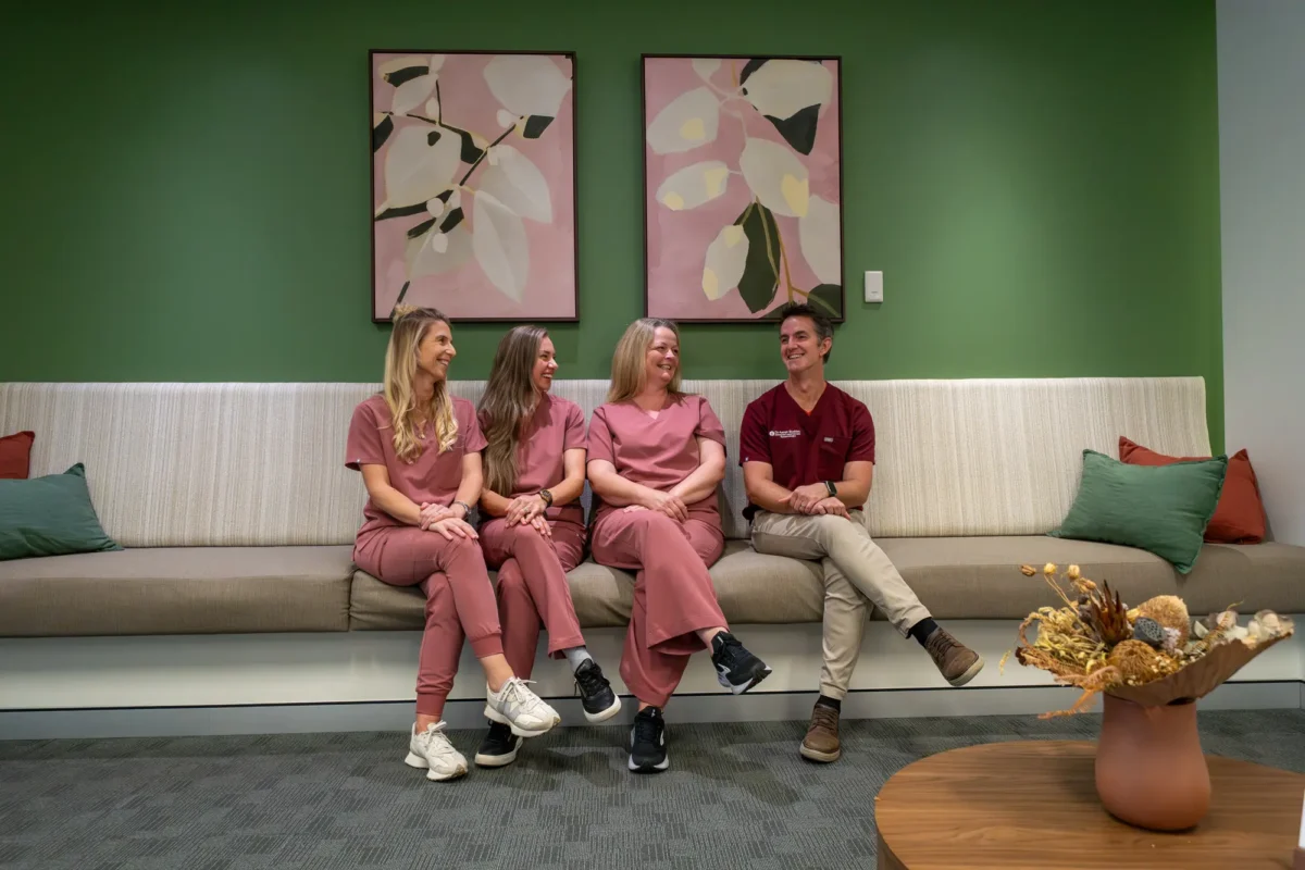
Take control of your gynaecological health. Contact the team at Saige Ultrasound today to schedule your specialised endometriosis assessment. You can call us directly on 02 6652 1109, email referral@saigeultrasound.com.au, submit a Request Callback form online, or click Book Now to secure your appointment instantly.
This blog is brought to you in collaboration with Dr Aaron Budden Gynaecologist from Saige Health & Wellness and Saige Ultrasound, and the independent radiology teams at Kare Radiology.
Further Links to learn more about Endometriosis
https://endometriosisaustralia.org/
https://www.jeanhailes.org.au/health-topics/endometriosis/
https://draaronbudden.com.au/conditions/pelvic-pain-endometriosis/
https://www.saigehealthandwellness.com.au/conditions/endometriosis-and-pelvic-pain
https://www.mayoclinic.org/diseases-conditions/endometriosis/symptoms-causes/syc-20354656
https://www.aihw.gov.au/reports/chronic-disease/endometriosis/contents/what-is-endometriosis