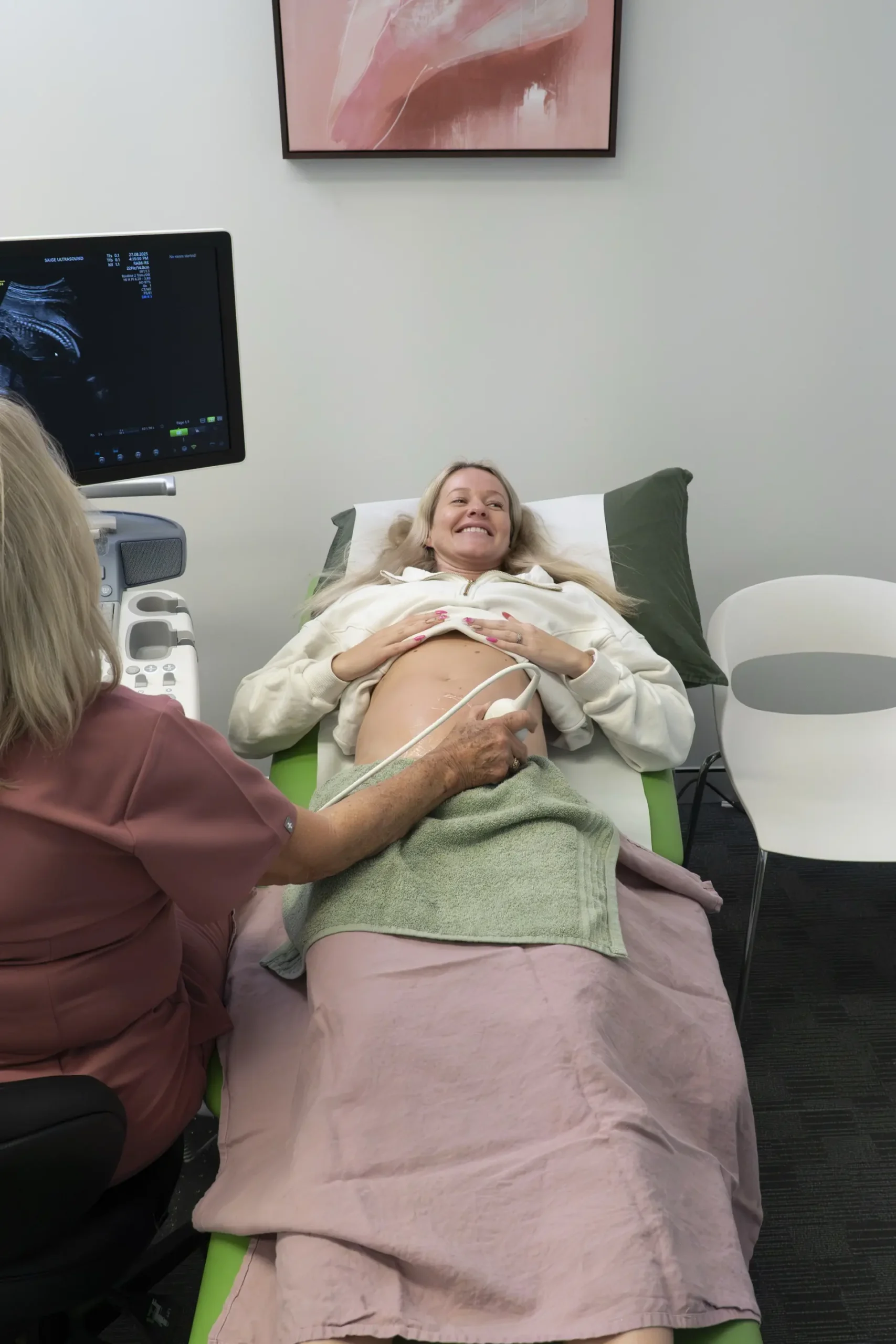
Coffs Coast’s only Obstetric & Gynaecological Specialist-led Ultrasound Clinic
PREGNANCY
Our pregnancy ultrasound services focus on monitoring fetal development, detecting any abnormalities, and ensuring the well-being of both mother and baby. We provide detailed reports and compassionate care throughout the process with specialist obstetricians available to talk through your result and provide advice on your next steps
What’s Involved
Undergoing ultrasound in pregnancy is one of the best ways to ensure you and your baby are growing well. But these days it’s also a great way to get to know your little one and honestly we’re here for it.
Our diagnostic services include confirmation of your dates, assessment of chromosomal and pre-eclampsia risk, structural assessments (known as a morphology), and checking for growth and placental location.
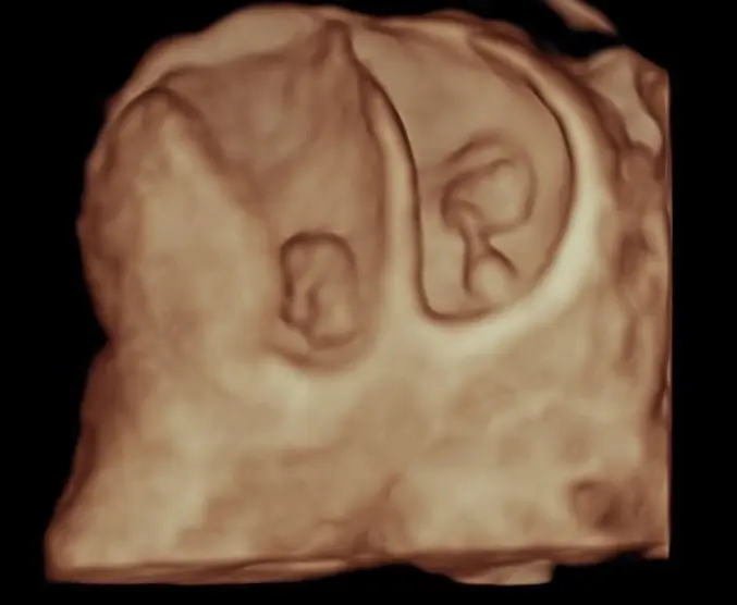
We also offer non-diagnostic 3D and 4D scans to help you and your family bond before your baby arrives.
Dating Scan
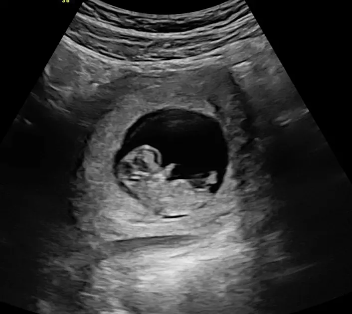
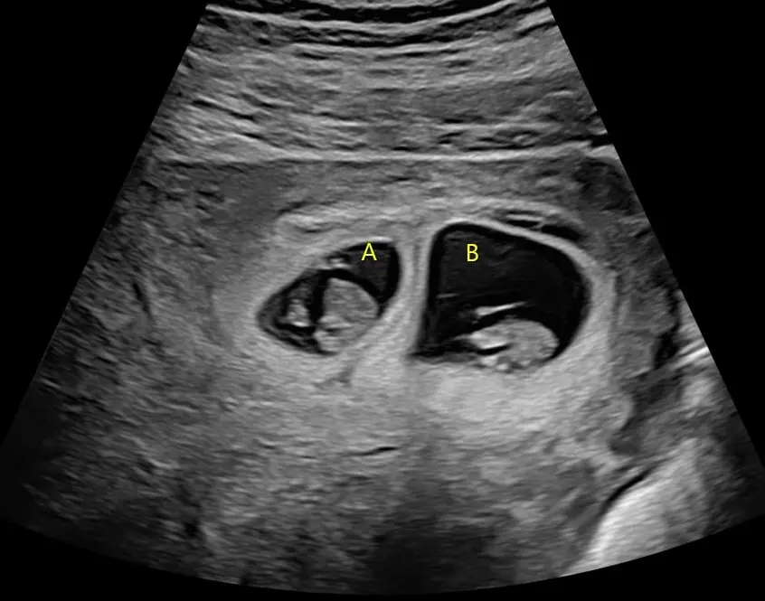
Whilst an early pregnancy ultrasound is not recommended for all pregnancies, we recognise it remains an important test for those with complications of early pregnancy, are unsure of their conception date, or just need extra support in these early days. It can help check the viability of your pregnancy, check the pregnancy is growing in the right place, and identify twins early.
The start of any pregnancy, whether it is planned or not, can be filled with questions and worries for everyone. The team are here to support you be it:
- Bleeding in early pregnancy
- Recurrent miscarriage
- Pregnancy of uncertain viability
- or just trying to work it out
1st Trimester
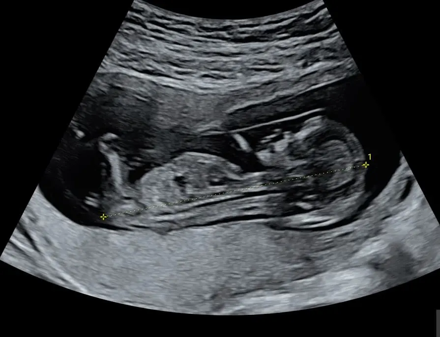
The days of considering the 12 week ultrasound as just a screening test for chromosome abnormalities (like Down syndrome) are long gone. Advances in ultrasound technology mean that we can start to examine your baby’s anatomy much sooner than in the past. The first trimester anatomy scan is performed between 12 and 14 weeks gestation and is an important option to check the development of your baby no matter your choices for chromosomal screening.
During this ultrasound we will check your baby’s heart beat, growth as well as the due date for your pregnancy. Our dedicated team will also check:
- Anatomy of your baby
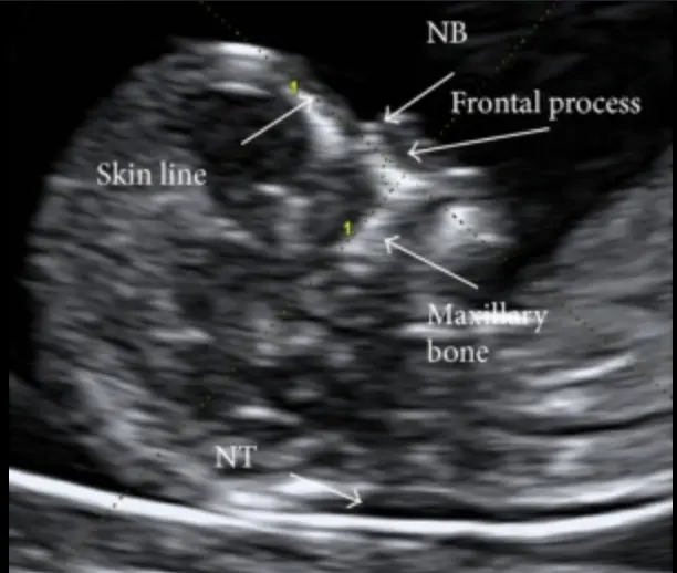
- Nuchal Translucency
- Placental location and ovaries
With advances in ultrasound technology, your baby’s anatomy can be seen in great detail between 12 and 14 weeks gestation. It is now possible to see your baby’s arms, legs, fingers, and toes as well as skull, heart, brain, stomach, and kidneys.
During this ultrasound we undertake additional tests to screen your baby (if requested) for you, your cervical length, and risks of developing pre-eclampsia. This involves:
- Chromosomal anomalies by measuring the nuchal translucency thickness
- Risk of pre-eclampsia by measuring the uterine artery blood flow
- Assess for pre-term birth through measuring the length of the cervix
Whether you choose to have screening for chromosomal abnormalities or not, the 1st trimester anatomy scan can provide much more information on your growing baby.
And if you have chosen to undertake NIPT, we welcome you for check of fetal viability on the day of your blood test.
Morphology
A fetal morphology ultrasound, also known as the 20-week scan or anatomy scan, is a detailed ultrasound examination that looks closely at your baby’s development and checks for any abnormalities. It is a detailed check of your baby from head to toe and is usually performed between 18 and 22 weeks of pregnancy.
This scan provides vital information about your baby’s health and development. It allows your healthcare provider to:
- Check the baby’s size and growth
- Examine the structure of the baby’s organs (such as the brain, heart, kidneys, spine, and limbs)
- Look at the placenta, amniotic fluid, and umbilical cord
- Confirm the position of the baby and placenta
- Assess for physical abnormalities or birth defects
- (If you wish) determine the baby’s sex
The procedure is performed using a transducer on your abdomen. It is a safe and non-invasive test that uses sound waves, not radiation, and poses no known risks to you or your baby. The scan typically takes around 45 to 60 minutes, but this may vary depending on the baby’s position and movement. Sometimes, if the baby is not in an ideal position to view certain structures, a follow-up scan may be scheduled.
3rd Trimester
A third trimester ultrasound is an ultrasound scan performed during the final stage of pregnancy, typically after 28 weeks. It is usually recommended when there is a medical reason to assess the baby’s wellbeing, growth, or position. Unlike the routine scans performed earlier in pregnancy, not everyone will need a third trimester ultrasound unless advised by their healthcare provider.
A third trimester ultrasound may be recommended if there are concerns such as reduced fetal movements, high or low levels of amniotic fluid, abnormal fundal height (the size of your belly), maternal medical conditions (like high blood pressure or diabetes), previous pregnancy complications, or if the baby is suspected to be too small or too large for gestational age.
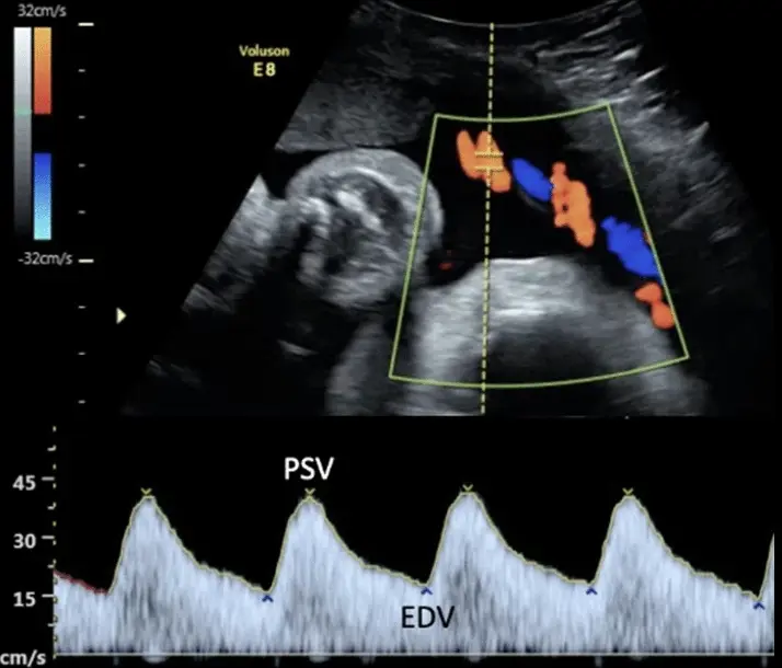
The main purpose of a third trimester ultrasound is to check how your baby is growing and developing. It allows the sonographer to measure your baby’s size, monitor growth over time, and assess the amount of amniotic fluid around the baby. The position of the baby (whether head-down, breech, or transverse) and the location and appearance of the placenta are also checked. Blood flow in the umbilical cord and other vessels may be assessed using Doppler ultrasound if there are concerns about the baby’s growth or overall health.
You do not usually need a full bladder for this scan, and no special preparation is required. The scan usually takes around 20 to 30 minutes, but this may vary depending on the baby’s position and the specific reason for the ultrasound. You can typically bring a partner or support person with you.
Baby Bonding
We connect to each other through our shared lived experiences. And pregnancy and birth are amongst the powerful experiences we can ever have. Utilising imaging technology enhances our own experiences further and also allows to share with our loved ones. Whilst we aim to capture great 3D images of your little one at every regular ultrasound, we also offer a dedicated ultrasound for bonding with your baby.
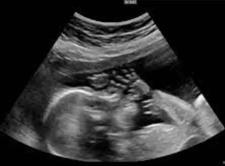
A 3D baby bonding ultrasound is a special type of non-medical ultrasound designed to give expectant parents a more detailed and lifelike view of their baby during pregnancy. Unlike standard diagnostic ultrasounds, which are used to assess your baby’s health and development, a 3D bonding scan focuses on creating clear, three-dimensional images that highlight your baby’s facial features, movements, and expressions in the womb. These ultrasounds are usually performed between 26 and 32 weeks of pregnancy, when there is enough fat under the baby’s skin to see the face clearly, but still enough amniotic fluid to produce quality images.
A 3D bonding ultrasound does not replace your routine medical ultrasounds and should be considered a complementary experience for emotional connection and enjoyment. It is important to have your standard medical ultrasounds completed as recommended by your healthcare provider to monitor your baby’s health. If the sonographer performing the 3D scan notices anything unusual, they will advise you to follow up with your doctor or midwife.