Coffs Coast’s only Obstetric & Gynaecological Specialist-led Ultrasound Clinic
GYNAECOLOGY
Expert diagnostic imaging to guide the best treatment options.
What’s Involved
When it comes to issues of gynaecology, ultrasound is certainly not a one size fits all kind of deal and we are here to ensure you and your doctors get the answers you need.
From basic ultrasounds for issues such as abnormal bleeding, specialised ultrasound for pain and fertility, through to ultrasound based procedures, we have you covered.
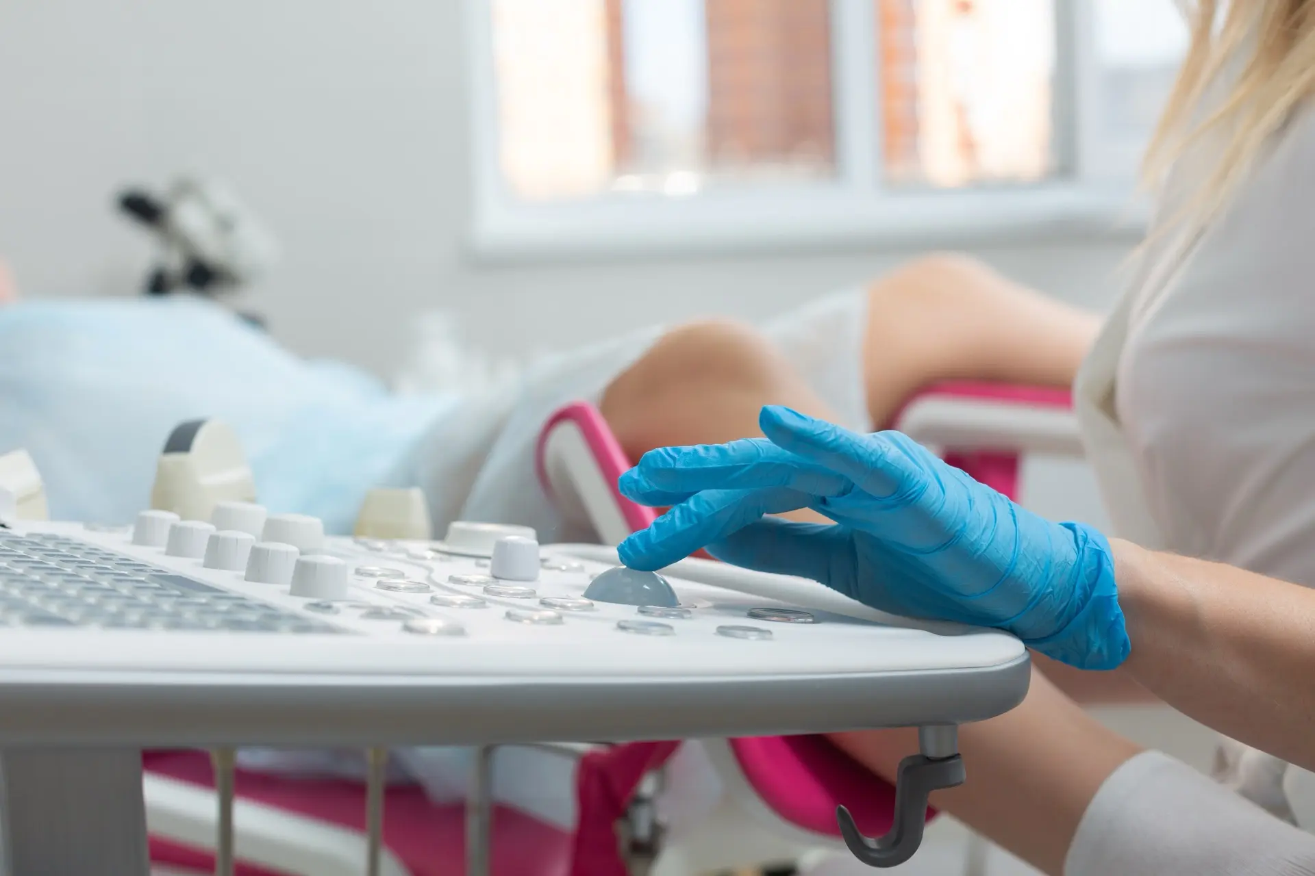
Traditional Pelvic Ultrasound
A standard pelvic ultrasound is a common and safe imaging test used to examine the organs and structures within the pelvis. It is often used to assess the uterus, ovaries, endometrium (lining of the uterus), cervix, bladder, and surrounding tissues
This type of ultrasound is frequently requested to investigate symptoms such as
- Abnormal bleeding including menopuse
- Heavy periods
- Painful periods
- Infertility
- Monitor known conditions like ovarian cysts or fibroids
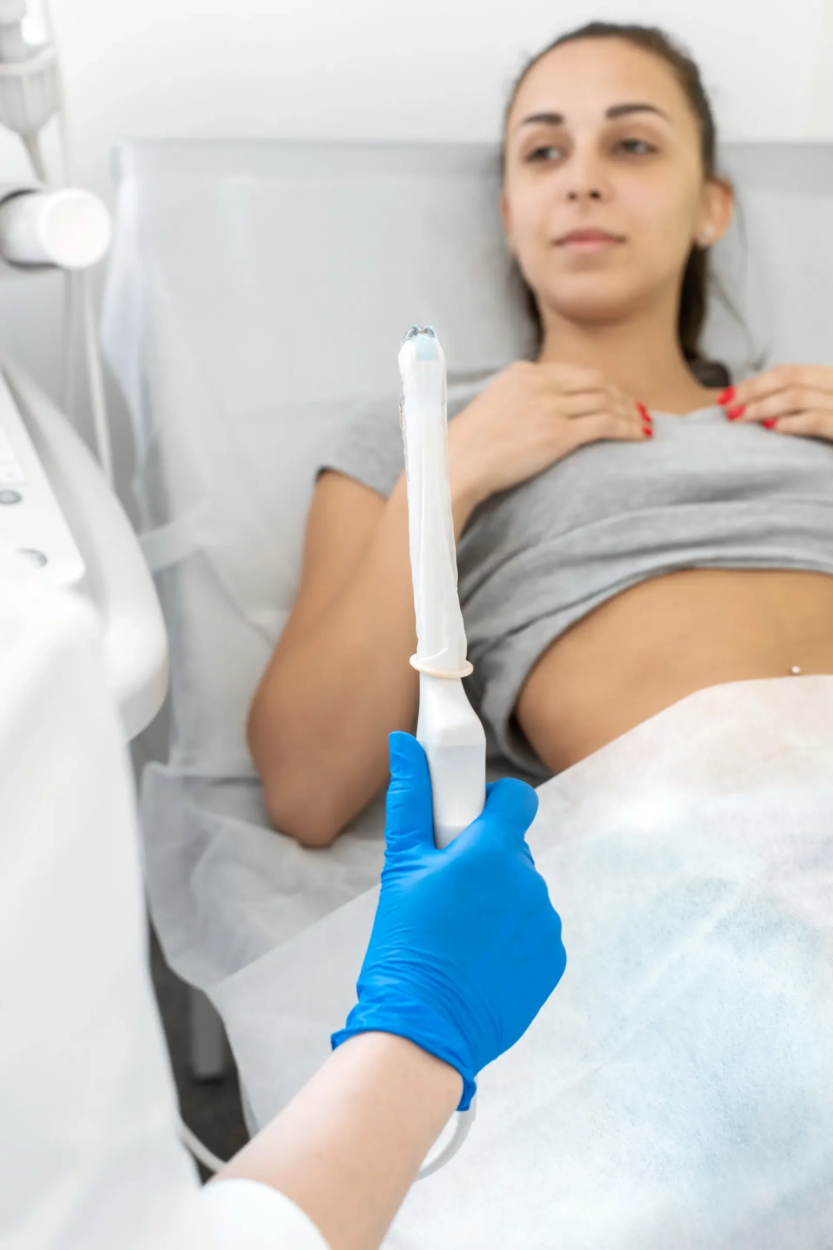
There are two main types of pelvic ultrasound: transabdominal and transvaginal. A transabdominal ultrasound is performed by placing a probe on the lower abdomen, and a full bladder is usually required to help lift the pelvic organs into better view. You may be asked to drink water and not empty your bladder before the scan. A transvaginal ultrasound involves gently inserting a specially designed probe into the vagina, which provides a closer and more detailed view of the pelvic organs. This part of the scan is usually done with an empty bladder and is only performed with your consent.
Deep Infiltrating Endometriosis
The deep infiltrating endometriosis (DIE) scan is a thorough assessment of the uterus, ovaries, bowel, bladder, ligaments, and surrounding structures for those with pelvic pain.
It requires transvaginal assessment for accurate diagnosis however we no longer require bowel preparation.
What is Deep Infiltrating Endometriosis?
Superficial endometriosis can be thought of as disease that is just on the surface. When endometriosis grows into tissues and organs, like ovaries and the bowel, we consider this as deep infiltrating disease.
What is involved in a DIE ultrasound?
A standard pelvic ultrasound will look at the uterus, ovaries, and the immediate surrounds (the adnexa). A DIE ultrasound looks at all these organs as well as looking at specific locations where endometriosis typically likes to develop such the anterior rectum, the ligaments that support the pelvis, the bladder, and fallopian tubes. It takes around twice as long as a normal pelvic ultrasound and requires an internal ultrasound to see the organs and tissues of the pelvis well.
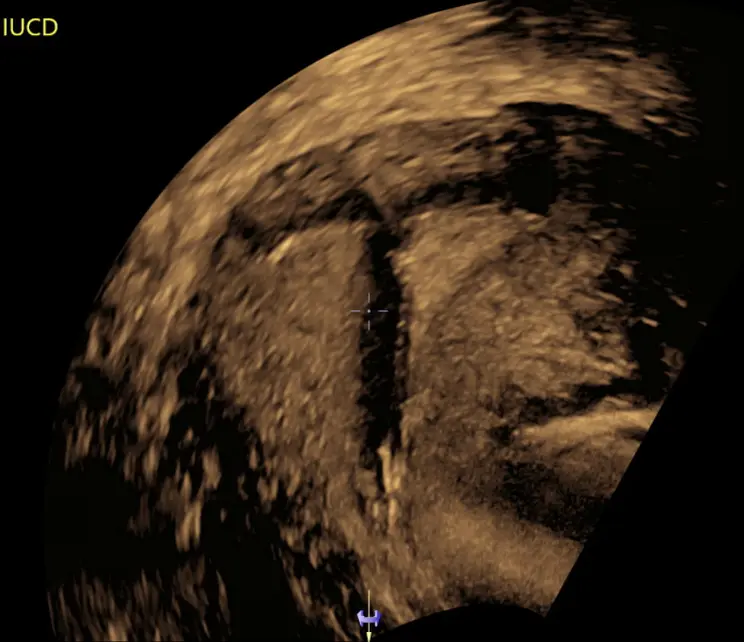
Placement, location, and removal of Intrauterine Device (e.g Mirena)
An Intrauterine Device (IUD) assessment, including 3D imaging, is a routine part of a gynaecological ultrasound examination.
When working correctly, an IUD should not not cause any unusual symptoms. IUD assessments are generally performed when a woman has symptoms that suggest a potential issue with her IUD (like pelvic pain or abnormal bleeding), or when a healthcare provider is unable to locate the strings of the IUD during a physical examination.
The main objectives of an IUD assessment are:
Presence of the IUD: IUDs have been known to leave the body without the woman noticing. An IUD assessment ultrasound can confirm whether the IUD is still present in the uterus.
Position of the IUD: The assessment verifies that the IUD is in the correct position within the uterus.
Complications: An IUD assessment ultrasound may be able to identify potential complications related to the IUD or its placement. This includes complications such as:
- Embedding into the uterine wall
- Perforation (where the IUD punctures the wall of the uterus)
At times an IUD requires removal but the strings are not clearly visible or brought into view with a string finder. When this occurs, it is recommended to undertake removal of the IUD under vision either by hysteroscopy (an operation where you will be under sedation or general anaesthetic) or under ultrasound guidance. At Saige Ultrasound we offer the removal of IUD under ultrasound guidance with the option of having methoxyflurane (green whistle) to improve comfort.
At different times, uterine anatomy (such as fibroids or a large caesarean scar) may cause difficulty to safely insert an IUD. Ultrasound guidance can reduce the risks of blind insertion of IUD in these circumstances and can ensure its adequate location and deployment prior to completion of the procedure. Similar to removal, Saige ultrasound offers the option of methoyxflurane to improve patient comfort.