Coffs Coast’s only Obstetric & Gynaecological Specialist-led Ultrasound Clinic
FERTILITY
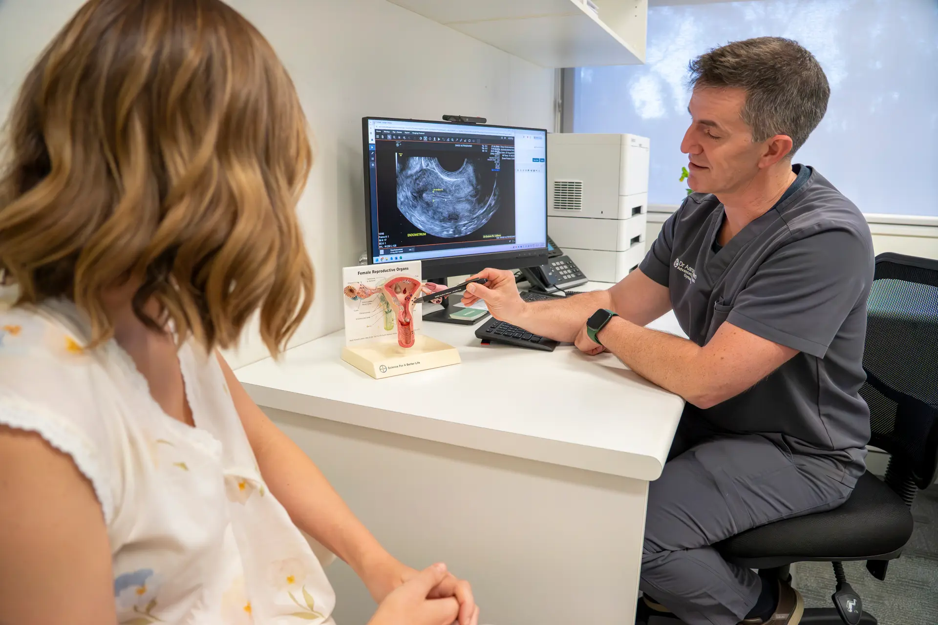
When it comes to difficulties in having a baby, ultrasound is crucial in both diagnosis of any anatomical abnormalities as well as providing ongoing assessment of the ovaries during ongoing cycles.
What’s Involved: Fertility workup and diagnosis
During the investigation for any fertility issues, your doctor will likely refer you for a routine pelvic ultrasound to rule out issues that may affect your fertility and approach to IVF. This may include:
- Polyps and Fibroids
- Caesarean scar gaps
- Overall size of the uterus
- Abnormalities of the ovary
The following components are commonly included in a routine gynaecological ultrasound:
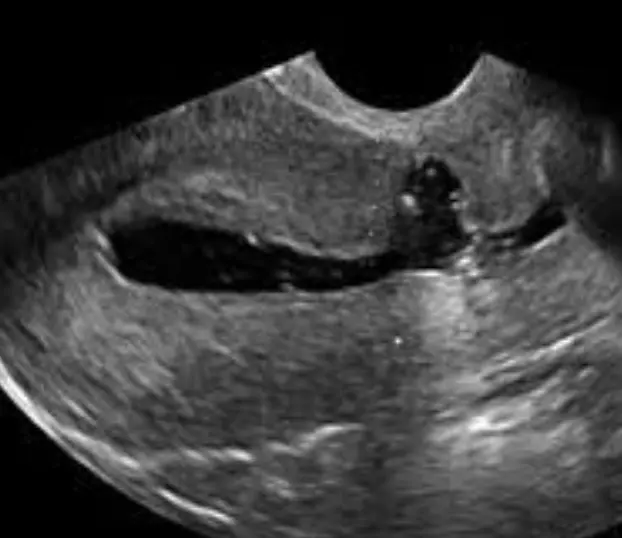
Uterus: We examine the size, position and shape of the uterus such as a septate or bicornuate uterus, and evaluate the endometrium (the lining of the uterus) for thickness and polyps. The uterus may also contain fibroid.
We also offer non-diagnostic 3D and 4D scans to help you and your family bond before your baby arrives.
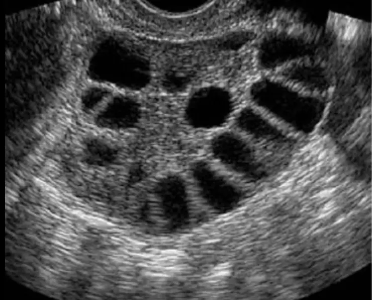
Ovaries: The scan assesses the size and presence of cysts. During the assessment, we will assess the number of antral follicles and follicle sizes.
Fallopian tubes: While these structures are usually not seen unless they are dilated or filled with fluid, you may also be recommended to have a test of tubal patency to check the connection from uterus to ovary.
This can be done at surgery or through a special ultrasound called High contrast sonography (HyCoSy)
Assessment for Endometriosis: an assessment for evidence of Endometriosis is performed in all gynaecological ultrasounds.
Saline Sonogram & HyCoSy
There are times when a routine pelvic ultrasound identifies an abnormality of the uterus but because the inside of the uterus is closed, it is hard to completely identify the limits of the abnormality.
This may include:
- Polyp size and location
- Fibroid size and location
- Significance of a caesarean scar gap in the uterine wall
Saline Sonography (SIS) is a specialised test used to understand the inside part (endometrial cavity). It involves a procedure to inject salt water (saline) into the uterine cavity whilst performing an ultrasound. It is used to detect abnormalities of the lining of the uterus. It is performed in conjunction with a transvaginal ultrasound to assess the pelvis.
You may be sent for a SIS to investigate:
- Fertility issues
- Recurrent miscarriage
- Bleeding issues such a postmenopausal bleeding or irregular bleeding
- Fibroids
- Polyps
The procedure needs to be performed between 3 to 9 days after the first day of your last menstrual period. If you have a very irregular cycle or infrequent periods it is advisable to contact us on day 1 of your period to make your booking.
The Sonographer or Doctor will explain the procedure to you and discuss your relevant history. A transvaginal ultrasound will be performed prior to the Saline Sonogram procedure.
The Doctor will insert a speculum into the vagina and apply an antiseptic solution to the cervix. A fine tube (2mm) will be passed through the cervix and into the uterine cavity. The speculum will then be removed and the transvaginal probe will be inserted. A solution of saline will be introduced into the uterus using the fine tube. You may feel a warm sensation and less frequently mild cramping similar to period pain. The Doctor will observe the solution pass through the uterine cavity and assess the lining of the uterus. When the procedure is finished the tube and probe will be removed.
High Contrast Sonography (HyCoSy) is an extension of the saline sonogram. HyCoSy is a specialised ultrasound procedure used to assess the inside of the uterus and the fallopian tubes. It involves inserting a solution of contrast material into the uterine cavity and observing the flow within the fallopian tubes. HyCoSy is used to detect blockages of the fallopian tubes and abnormalities of the uterus.
Our specialist team undertake these procedures with the best care whilst limiting the discomfort as much as possible.
Ovulation Tracking
One of ultrasounds best abilities is to look at how your eggs are growing. This may be done for
- IVF cycles
- During ovulation induction and IUI
- Under “track and try” cycles
It involves an internal ultrasound to measure the number and size of the follicles in each ovary. The first scan will often occur several days before ovulation is expected and continue every 2-3 days until the follicles (eggs) are the correct size for your specific fertility treatment.