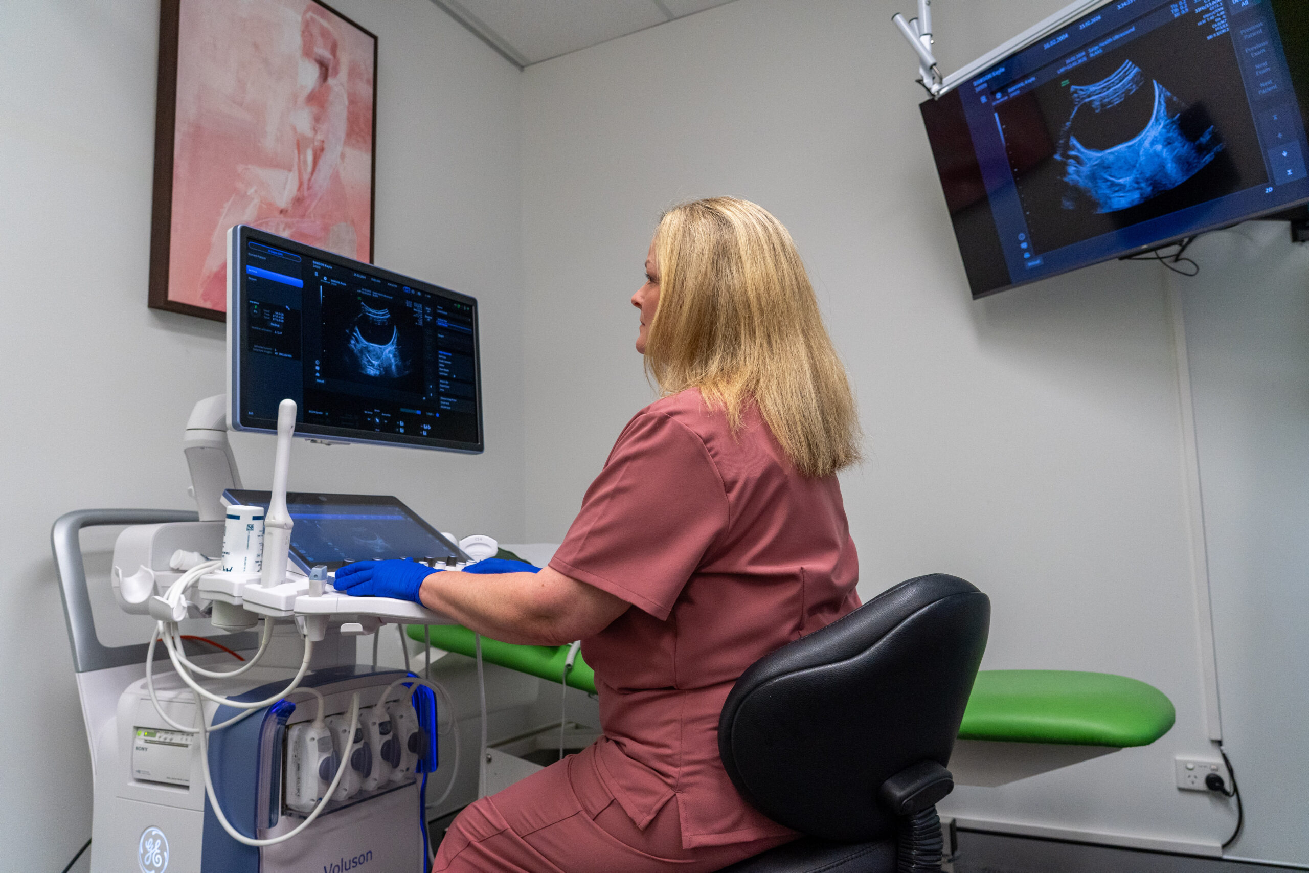
If you’re someone who’s lived with chronic pelvic pain, heavy bleeding, or fertility struggles with no real answers, there can be nothing more disheartening for you to hear than: “Good news, your ultrasound came back completely clear.”
You might be thinking, “But isn’t this a good thing?” While an all-clear result should bring a sense of relief, for many women, it only deepens the confusion and frustration even more. When you know your body, and you know your pain is real, but the imaging report in your hands suggests everything is normal, you know deep down that something isn’t adding up.
The truth is that a standard pelvic ultrasound is primarily designed to look for broad, structural issues, such as large ovarian cysts or obvious fibroids. It is rarely equipped to detect subtle adhesions, adenomyosis, or Deep Infiltrating Endometriosis (DIE). However, it is still common practice to send most women for a standard pelvic ultrasound despite their symptoms, and this is why misdiagnosis rates remain at an all-time high.
Thankfully at Saige Ultrasound, we intentionally do things differently. We believe that getting accurate diagnoses and clarity for patients requires more than basic equipment. It requires specialised scanning protocols and a dedicated obstetric & gynaecological team involved in your care throughout your entire journey.
The Saige Difference: Real-Time Specialist Care
In a typical radiology environment, your scan will be performed by a general sonographer, and your static images are sent off to an offsite doctor or radiologist with no prior connection to you to perform the assessment of the images. They evaluate snapshots without knowing your full clinical history, seeing your physical responses, or understanding where your pain is localised, providing their analysis based solely off the scanned images.
Saige Ultrasound is a specialist-led model that is unique to the Coffs Coast and Mid North Coast of NSW. We triage every referral up front so your scan is tailored to your exact clinical history. For complex cases, including patients with previous surgeries, ongoing severe pain, or specific bowel symptoms, scans are scheduled so our obstetrician and gynaecologist specialist, Dr Aaron Budden, can be in the room alongside our expert sonographers, asking questions and evaluating findings as the scan happens.
For other scans, Dr Aaron provides dedicated on-site review and reporting. He works closely with the sonographer to analyse what was captured on the screen, what was physically felt during the exam, and any physical pain triggers.
Having a practising specialist evaluating organ mobility, tissue sliding, and subtle anatomical markers in real time turns a routine imaging appointment into an active, comprehensive clinical assessment. This collaborative approach helps our team highlight complex conditions that basic scans can’t show and reduces the chances of you receiving the “your ultrasound came back clear” result.
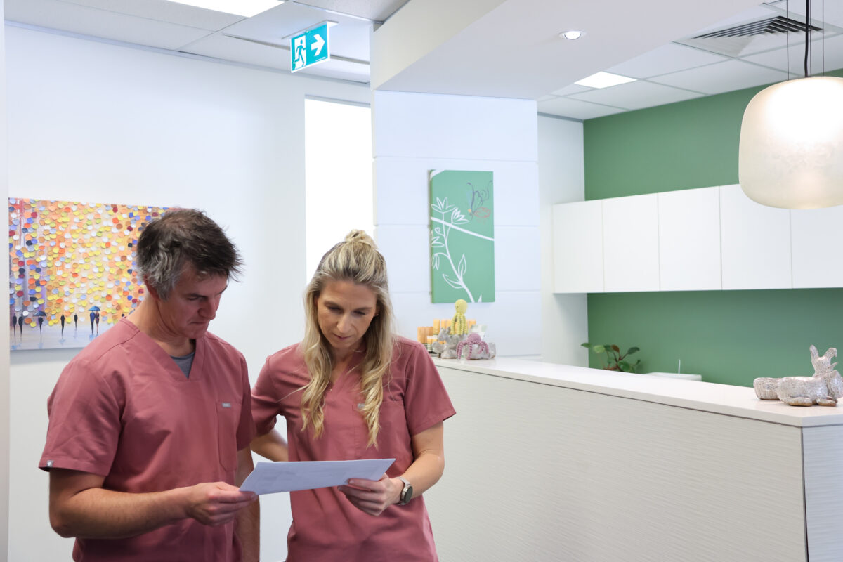
Which Scan is Right For Me? Standard vs. Complex vs. DIE Mapping
A basic overview is quite often not enough to explain what is happening inside your pelvis. Thankfully, with more medical advances, we now have access to different types of gynaecological scans that we can utilise, depending on your clinical history and symptoms. These can help ensure you receive the right information, the correct diagnosis and the clarity you need. The types of ultrasound scans available fall into three distinct levels, which are explained below:
1. Standard Pelvic Ultrasound (Basic Screening)
- What it is: A foundational scan of the pelvic organs, typically using a combination of transabdominal and transvaginal ultrasound techniques.
- What it looks for: Major anatomical structures, large ovarian cysts, prominent uterine fibroids, endometrial thickness, and overall organ positioning.
- When it’s used: Excellent for routine check-ups, investigating sudden acute pelvic pain, or checking early pregnancy viability.
- Why it can fall short: A standard scan is a visual snapshot. It checks if organs are present and of normal size, but it rarely tests if those organs are stuck together by scar tissue or inflamed by microscopic lesions, which is often a big factor in some gynaecological problems.
2. Complex Gynaecological Ultrasound (Targeted Clinical Evaluation)
- What it is: A specialised assessment designed to investigate specific, ongoing gynaecological symptoms such as severe dysmenorrhea (painful periods), heavy menstrual bleeding, or unexplained fertility challenges.
- What it looks for: Subtle uterine pathologies like adenomyosis (where endometrial tissue grows into the muscular wall of the uterus), exact mapping of fibroid locations (submucosal, intramural, or subserosal), detailed ovarian cyst classification (distinguishing between simple fluid cysts, dermoids, and endometriomas/chocolate cysts), and initial organ mobility tests.
- The Clinical Edge: When specialists like Dr Aaron can understand surgical and fertility impacts, getting a complex scan with the specialist can evaluate how these conditions affect organ function, uterine lining quality, and reproductive pathways.
3. Deep Infiltrating Endometriosis (DIE) Mapping Scan (Advanced Protocols)
- What it is: A highly specialised, extended imaging protocol specifically designed to detect and map deep endometriosis nodules and pelvic adhesions. This scan requires advanced training, high-resolution equipment, and specialised dynamic maneuvers.
- What it looks for: Deep endometriotic lesions that extend beneath the peritoneum (the lining of the pelvis). This includes assessing:
- The Sliding Sign: Using gentle, controlled pressure to check if the uterus and ovaries slide freely against the bowel and pelvic walls, or if they are “frozen” in place by adhesions.
- Pouch of Douglas: Checking for obliteration or scar tissue behind the uterus.
- Bowel & Bladder Involvement: Identifying nodules on the rectosigmoid bowel wall, bladder base, or ureters.
- Uterosacral Ligaments & Vaginal Vault: Locating deep, painful nodules that frequently cause pain during intercourse (dyspareunia).
- Why it matters: A DIE mapping scan creates a detailed “road map” of your pelvis. If surgery is a requirement down the track, your surgeon knows exactly where the disease is located before making an incision, significantly improving surgical safety and outcomes.
Want a quick reference guide to bring to your doctor? Ask for our Endometriosis Diagnostic Guide
to explore these 3 scan pathways in detail and see how to prepare for your appointment.
A Report That Provides Real Clarity
A standard radiology report often provides a brief, inconclusive line like “Uterus and ovaries appear within normal limits.” When general practitioners (GPs) receive basic reports with little direction or guidance like this, it makes it hard for them to put actionable and accurate steps in place to help provide relief for patients suffering from debilitating symptoms. This is commonly why patients are left feeling frustrated and unvalidated.
When you have your scan done through Saige Ultrasound, your scans are interpreted by specialists, with the report sent back to your referring GP, providing detailed, precise, and actionable results. Rather than generic observations, Saige Ultrasound provides diagnostic breakdowns that map out exact disease locations, organ mobility scores, and clinical implications to help patients on their journey to a resolution.
This detailed feedback we provide gives your GP a clear roadmap so they can:
- Confidently confirm or rule out complex conditions without requiring immediate exploratory surgery.
- Tailor medical management plans (such as hormonal therapies or pain management) based on exact disease severity.
- Facilitate precise, direct referrals to a gynaecological team when advanced intervention is needed.
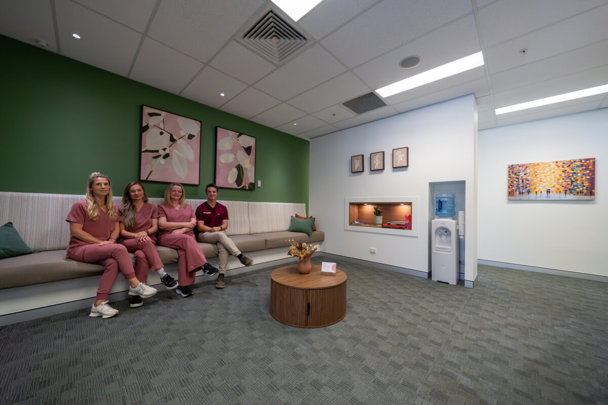
You Deserve Clear Answers
If you are living with unexplained pelvic pain, heavy periods, fertility hurdles or other gynaecological symptoms, a “clear” scan should not be the end of your diagnostic journey. When you have a team supporting you who are able to provide specialist oversight and advanced scanning protocols, getting clear, accurate answers is possible. Before giving up, talk to your GP and push for further support and more clarity.
Request a specialist-led pelvic ultrasound with Saige Ultrasound, or call us directly on 02 6652 1109, email referral@saigeultrasound.com.au, submit a Request Callback form online, or click Book Now to secure your appointment instantly.